 Instructional Biology
Protein medicines are long chains of amino acids folded into precise shapes, and most proteins are only marginally stable in that folded state. When the fold opens, even partly, the exposed chains tend to stick to one another, and the clumps they form can be extremely stable and long-lived [1]. This process, protein aggregation in biopharmaceuticals, is one of the central quality problems in making biologic drugs. A review in the International Journal of Pharmaceutics called it arguably the most common and troubling form of protein instability, met in almost every stage of protein drug development [2].
This article follows a biologic from cell culture to the patient: where aggregates form, why regulators and clinicians care, and how the industry measures and controls them. For the basic physics of unfolding, start with our explainer Why Does Heat Denature Proteins?
What is protein aggregation in biopharmaceuticals?
In a biologic, aggregation means protein molecules assembling into stable complexes of two or more units; each single unit is called the monomer [1]. The aggregates that matter most are held together by strong contacts and need some unfolding or misfolding to form, because a partly opened protein exposes stretches of amino acids that bind tightly to neighboring chains [1]. These "hot spots" tend to be hydrophobic, uncharged and prone to forming beta sheets [1]. Once formed, such aggregates are very difficult to reverse: they do not come apart to any significant degree on dilution or a shift in pH [1].
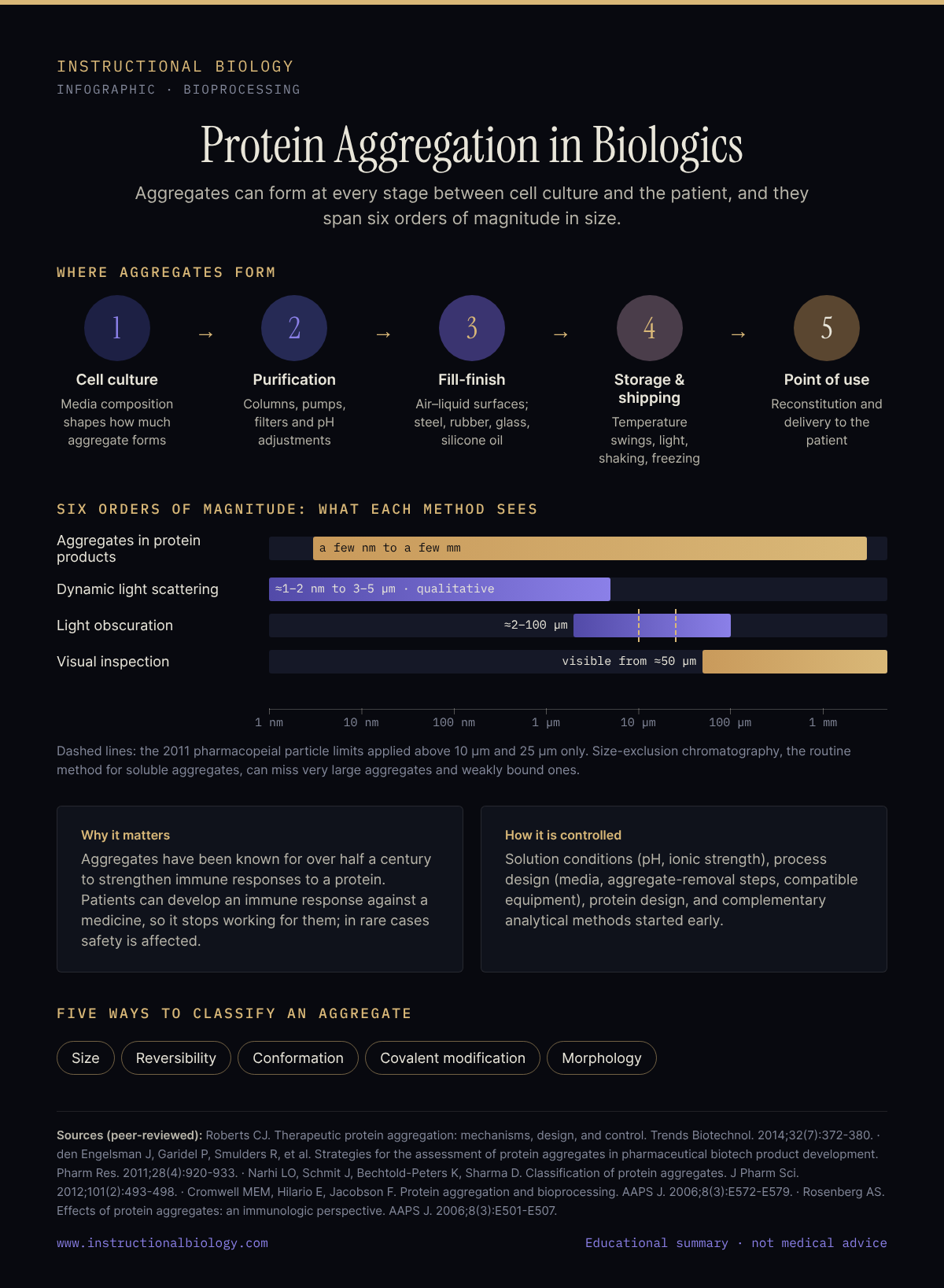
Aggregates also come in a very wide range of sizes, from two-molecule dimers to particles visible to the eye. The range spans up to six orders of magnitude, from a few nanometers to a few millimeters [3]. To make results comparable between laboratories, Narhi and colleagues proposed describing every aggregate on five independent axes: size, reversibility, conformation, covalent modification and morphology [4].
Where aggregates form: from cell culture to the syringe
Aggregation is not one event at one step. It can begin during production and continue through storage, shipment and delivery to the patient [3], and reviews of its immune effects describe the same risk across manufacturing, shipping and storage [5]. The main stress points:
- Cell culture. The amount of aggregate made during cell culture and purification can be influenced by carefully controlling the environment, including the components of the growth media [6].
- Purification. Contact with chromatography columns, pumps, pipes, vessels and filters, and pH adjustments along the way, can all induce aggregation [3].
- Fill-finish. Mixing and filling create air–liquid and liquid–solid interfaces. Foreign particles can seed aggregates too: stainless steel from filling pumps, rubber from stoppers, glass from heated containers and silicone oil droplets from siliconized syringes [3].
- Storage and shipping. Temperature swings, light, shaking and contact with ice during freezing are recognized triggers [3]. Even a product that leaves purification as high-purity monomer will, in most if not all cases, form some irreversible aggregate over time as it is stored and transported. Under well-chosen conditions those rates are low, which is what allows target shelf lives of two years or more [1].
- At the point of use. Reconstituting a freeze-dried product creates fresh interfaces, and aggregates can also form during delivery to the patient [3].
Different stresses leave different chemical fingerprints. In an Amgen study of an IgG2 antibody, aggregates made by mechanical stress carried methionine and/or tryptophan oxidation, consistent with damage at the air–liquid interface, while heat-induced aggregates showed significant deamidation and almost no oxidation [7]. Each stress follows its own pathway, which is one reason aggregates cannot be treated as a single interchangeable impurity.
Why aggregates matter: immunogenicity
The ability of protein aggregates to strengthen immune responses against the single-molecule form of a protein has been known for over half a century [8]. Aggregates were linked to antibody-mediated adverse events with early therapeutic protein products, including intravenous immune globulin and human growth hormone [8]. Proteins displayed in a highly arrayed structure, as in large non-denatured aggregates, are highly potent at inducing antibody responses even without T-cell help, possibly because they cross-link many B-cell receptors at once [8].
For a patient, the consequence can be an immune response against the medicine, so that the drug no longer works for them; in rare cases it can also cause serious safety issues [1]. In extreme and rare cases, patients have developed immunity to their own matching protein, as in pure red cell aplasia [1]. Aggregation can raise this risk even for humanized and fully human proteins [1].
Which aggregate species drive these responses is still not settled. A 2016 review points to gaps in understanding which physical and chemical features trigger immunity, and to a lack of standardized methods for measuring them [5]. Because of that uncertainty, regulators err on the side of caution and expect the amount and properties of all detectable aggregates to be well controlled, reproducible and monitored [1]. Current evidence from animal models suggests that smaller aggregates and sub-visible particles are of greater concern than large insoluble clumps [1].
How protein aggregates are measured
No single instrument covers six orders of magnitude, so developers combine complementary methods, each with its own blind spots [3]:
- Size-exclusion chromatography (SEC) is the routine quality-control method for soluble aggregates. Very large aggregates can be filtered out by the column and missed, and weakly associated ones can fall apart during the run [3].
- SDS-PAGE and capillary electrophoresis (CE-SDS) detect covalent aggregates only, because the detergent breaks non-covalent ones apart [3].
- Dynamic light scattering sizes particles from about 1–2 nm to 3–5 µm and can detect trace amounts of large species below 0.01% w/v, but its results are qualitative rather than quantitative [3].
- Analytical ultracentrifugation characterizes aggregates in the formulation itself; for antibodies its quantification limit is about 1% at best, so it works best as a cross-check [3].
- Light obscuration, a pharmacopeial method, counts particles of roughly 2–100 µm. In the 2011 review, the compendial acceptance criteria applied only to particles larger than 10 and 25 µm; particles between 0.1 and 10 µm were not covered [3].
- Visual inspection catches visible particles, with a visibility threshold of about 50 µm for a spherical particle [3].
How protein aggregation is controlled
Control works on four levels: the solution, the process, the molecule and the analytical plan.
- Solution conditions. For several antibodies and other proteins, raising the pH well above the protein's isoelectric point or lowering ionic strength makes aggregates grow less easily, because the molecules repel one another more strongly [1]. Very few protein drugs are processed or stored at neutral pH and blood-like salt levels, so predictions made for physiological conditions are of questionable relevance to many protein medicines [1].
- Process design. Careful control of media and purification conditions limits how much aggregate forms, steps that remove aggregates have been used successfully at manufacturing scale, and equipment should be checked for compatibility with each protein [6]. Packaging, such as silicone-coated syringes, and leachables from glass or tubing should be tested for their effect on the product [3].
- Protein design. Engineers screen large libraries of variants, compare sequences with related proteins from organisms that evolved to tolerate aggregating stresses, or apply mechanistic knowledge: stabilizing the folded state, making protein surfaces more mutually repulsive, or removing aggregation hot spots [1].
- Analytical strategy. A practical plan pairs robust routine methods (SEC, SDS-PAGE or CE-SDS, light obscuration) with case-by-case extended characterization (analytical ultracentrifugation, field-flow fractionation, light scattering, microscopy), started early in development [3].
Our research direction
Our thesis at Instructional Biology is that aggregation control should begin upstream: if a protein is never pushed past the point where its fold begins to open, fewer molecules reach the unfolded states from which irreversible aggregation proceeds. That idea shapes our research on lower-temperature, native-state biomanufacturing and on the thermosensitive peptidome, the signaling peptides whose three-dimensional fold carries their function. This work is research-stage.
Sources
- Roberts CJ. Therapeutic protein aggregation: mechanisms, design, and control. Trends Biotechnol. 2014;32(7):372-380. doi:10.1016/j.tibtech.2014.05.005 · PMID 24908382
- Wang W. Protein aggregation and its inhibition in biopharmaceutics. Int J Pharm. 2005;289(1-2):1-30. doi:10.1016/j.ijpharm.2004.11.014 · PMID 15652195
- den Engelsman J, Garidel P, Smulders R, et al. Strategies for the assessment of protein aggregates in pharmaceutical biotech product development. Pharm Res. 2011;28(4):920-933. doi:10.1007/s11095-010-0297-1 · PMID 20972611
- Narhi LO, Schmit J, Bechtold-Peters K, Sharma D. Classification of protein aggregates. J Pharm Sci. 2012;101(2):493-498. doi:10.1002/jps.22790 · PMID 21989781
- Moussa EM, Panchal JP, Moorthy BS, et al. Immunogenicity of therapeutic protein aggregates. J Pharm Sci. 2016;105(2):417-430. doi:10.1016/j.xphs.2015.11.002 · PMID 26869409
- Cromwell MEM, Hilario E, Jacobson F. Protein aggregation and bioprocessing. AAPS J. 2006;8(3):E572-E579. doi:10.1208/aapsj080366 · PMID 17025275
- Luo Q, Joubert MK, Stevenson R, et al. Chemical modifications in therapeutic protein aggregates generated under different stress conditions. J Biol Chem. 2011;286(28):25134-25144. doi:10.1074/jbc.M110.160440 · PMID 21518762
- Rosenberg AS. Effects of protein aggregates: an immunologic perspective. AAPS J. 2006;8(3):E501-E507. doi:10.1208/aapsj080359 · PMID 17025268